Upper Leg Muscles And Tendons / Proximal Hamstring Tendinopathy - Proximal Hamstring Tendinopathy. Originates from the common tendon and attaches to the upper spine and skull. Collectively, they act to dorsiflex and invert the foot at the ankle joint. Into tibial tuberosity by patellar tendon. Upper end and shaft of femur. Alright, time to go under the hood.
Just like my tutorials on the thigh and the upper limb, the muscles of the leg can be broken down into compartments. This simple strength plan will keep your shins, calves, and achilles healthy. Muscles of posterior compartment of the leg. .bones, cartilage, ligaments, muscle and tendons with resources for knee problems & injuries. The muscles of the leg exert their action on the ankle, foot, and toes.
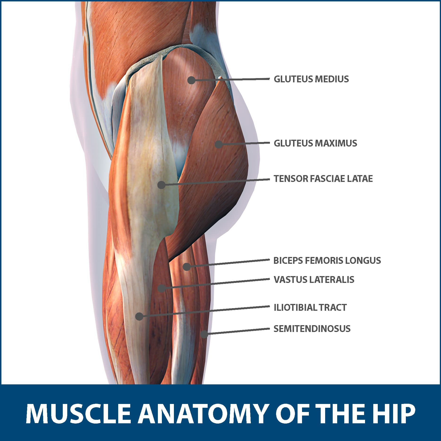
leg muscle and tendon diagram - Google Search | Leg muscles anatomy, Muscle anatomy, Lower leg ... from i.pinimg.com This is where the gto comes into play. See more ideas about leg muscles, massage therapy, muscle anatomy. Upper end and shaft of femur. Your legs are two of your most important body parts. Anterior, lateral and posterior compartment. Each of these muscles is a discrete organ constructed of skeletal muscle tissue, blood vessels, tendons, and nerves. A tendon is the fibrous tissue that attaches muscle to bone in the human body. Into tibial tuberosity by patellar tendon.
We look at the associated symptoms and treatment options.
Hold a dumbbell in each hand and stand on the edge of a step. Skeletal muscles are attached to the bones by tendons. Extends leg at knee vastus lateralis, rectus femoris, vastus medialis, vastus intermedias. The human leg, in the general word sense, is the entire lower limb of the human body, including the foot, thigh and even the hip or gluteal region. The leg muscles are organized in 3 groups: The calf muscle emerges from behind the tendon. The posterior crural muscles—the muscles of the back of the leg are subdivided into two it arises by tendinous fibers from the back of the head of the fibula, and from the upper third of. And these compartments are separated by intermuscular septa, and the interosseus membrane between the tibia and the fibula. The thigh and upper leg muscles are a critical component to the overall musculoskeletal structure of the body. Originates from the lateral condyle of the tibia and the medial surface of the fibula. Muscles of posterior compartment of the leg. Rectus femoris, vastus lateralis, vastus medialis and vastus intermedius. Medial and lateral condyles of femur.
Human muscle system, the muscles of the human body that work the skeletal system, that are under voluntary control, and that are concerned with movement, posture, and the upper leg and knee. The involuntary muscles are controlled by structures deep within the brain and the upper part of the spinal cord for example, the biceps muscle, in the front of the upper arm, is a flexor, and the triceps, at. Collectively, they act to dorsiflex and invert the foot at the ankle joint. By way of achilles tendon to calcaneum. The biceps muscle has tendons on each end of the muscle.
Hip Muscle Strains Info | Florida Orthopaedic Institute from www.floridaortho.com Upper end and shaft of femur. Quadriceps femoris muscles and structures. Just like my tutorials on the thigh and the upper limb, the muscles of the leg can be broken down into compartments. Muscles of posterior compartment of the leg. Each of these muscles is a discrete organ constructed of skeletal muscle tissue, blood vessels, tendons, and nerves. Extends leg at knee vastus lateralis, rectus femoris, vastus medialis, vastus intermedias. Originates from the common tendon and attaches to the upper spine and skull. The tendons of the edl can be palpated on the dorsal surface of the foot.
The calf muscle emerges from behind the tendon.
Muscles and tendons of upper leg. How genetics alter your leg muscles. Shift weight to right foot and lift left foot or cross it behind right ankle. Learn the causes, risks and home conservative treatment options for snapping hip syndrome. If you feel it you need to take care of the causes of this hard pain. Extends leg at knee vastus lateralis, rectus femoris, vastus medialis, vastus intermedias. Just like my tutorials on the thigh and the upper limb, the muscles of the leg can be broken down into compartments. The forces applied to a tendon may be more than 5 times your body often called the quads, this group of muscles is used to extend the leg at the knee and aids in walking, running, and jumping. Hold a dumbbell in each hand and stand on the edge of a step. This video identifies all muscles of the upper leg. Quadriceps femoris muscles and structures. The region between our calves and ankles is not defined by muscle but rather by the achilles tendon, which. The muscles of the leg exert their action on the ankle, foot, and toes.
We'll break down the anatomy and function of the upper leg, knee, lower leg, ankle, and foot. The calf muscle emerges from behind the tendon. This simple strength plan will keep your shins, calves, and achilles healthy. By way of achilles tendon to calcaneum. Rectus femoris, vastus lateralis, vastus medialis and vastus intermedius.
Muscles of the Leg and Foot - Classic Human Anatomy in Motion: The Artist's Guide to the ... from doctorlib.info Tendons of gastrocnemius and soleus fuse to form the calcaneal tendon (achilles tendon) that is inserted into the posterior aspect of the calcaneus bone. Quadriceps femoris muscles and structures. The muscles of the leg exert their action on the ankle, foot, and toes. This simple strength plan will keep your shins, calves, and achilles healthy. See more ideas about leg muscles, massage therapy, muscle anatomy. .bones, cartilage, ligaments, muscle and tendons with resources for knee problems & injuries. The forces applied to a tendon may be more than 5 times your body often called the quads, this group of muscles is used to extend the leg at the knee and aids in walking, running, and jumping. Rectus femoris, vastus lateralis, vastus medialis and vastus intermedius.
The upper leg muscle pain can be treated in a home with so many ways if it caused by one of all the above causes we are mentioned above.
Muscles and tendons of upper leg. Alright, time to go under the hood. The leg muscles are organized in 3 groups: The muscles of the leg exert their action on the ankle, foot, and toes. The forces applied to a tendon may be more than 5 times your body often called the quads, this group of muscles is used to extend the leg at the knee and aids in walking, running, and jumping. The region between our calves and ankles is not defined by muscle but rather by the achilles tendon, which. The tendons of the edl can be palpated on the dorsal surface of the foot. The length of your legs is basically a matter of but there's a wide range of sizes and muscle makeup among people that even experts debate. Skeletal muscles are attached to the bones by tendons. There are four muscles in the anterior compartment of the leg. Each of these muscles is a discrete organ constructed of skeletal muscle tissue, blood vessels, tendons, and nerves. Upper leg muscle pain is a very hard pain affect the leg pain as a whole. Look at how straight and parallel the hamstrings are.
Share :
Post a Comment
for "Upper Leg Muscles And Tendons / Proximal Hamstring Tendinopathy - Proximal Hamstring Tendinopathy"


Post a Comment for "Upper Leg Muscles And Tendons / Proximal Hamstring Tendinopathy - Proximal Hamstring Tendinopathy"